1880 Sismet Road Mississauga, ON L4W 1W9, Canada

EPIRETINAL MEMBRANE

An epiretinal membrane (ERM) is a layer of scar tissue that forms on the surface of the retina. It arises from a variety of ocular conditions including aging. Over time, an epiretinal membrane can cause the progressive wrinkling of the retinal surface, leading to severe and sometimes irreversible distortion and loss of sight. While surgery can be used to remove the epiretinal membrane and thus stop it from structurally changing the retina, it cannot reverse any severe damage done to the retina. Therefore, vision distorted or lost from the formation of an epiretinal membrane is sometimes impossible to restore. For the most part however, the removal of the epiretinal membrane is sufficient to clear the patient’s vision.

WHAT IS THE DISEASE?

As stated in the introduction, an epiretinal membrane is a layer of scar tissue that forms on the surface of the retina. It can lead to the development of cysts, bumps, or blisters in the retina. An epiretinal layer can arise from aging, an idiopathic disease, trauma, ocular inflammation, diabetes, or retinal detachment. Most often, ERM is predominately caused by aging, making it a widespread disease.

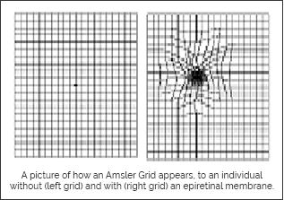
While the underlying cause for the initial formation of the ERM, such as trauma or chronic inflammation, can be stabilized, the ERM can continue to progress and lead to permanent distortion of the retina. It is through the structural alteration of the retina that epiretinal membranes cause severe distortion and loss of sight. Many patients complain of reduce clarity for fine acuity tasks such as reading or distortion of straight lines at a distance. Fortunately, this disease never leads to full blindness.

In addition to visual loss and distortion, an epiretinal membrane can cause straight objects to appear “wiggled”, especially at a distance. This effect becomes very clear with the use of an Amsler Grid, which checks whether the patient has a “wrinkled” retina, and thus “wiggly” vision.

PREVENTATION

Unfortunately, little can be done to prevent most epiretinal membrane formations, especially given that the majority is age-related. Protecting the eye from blunt trauma can reduce the incidence. Trauma can induce the formation of retinal tears and breaks which release retinal pigment epithelial (RPE) cells that accumulate in the central macular region of the eye. It is the unfortunate central placement of these cells that creates the noticeable distortion in vision. Certain ocular conditions precipitate the formation of these membranes including diabetic retinopathy.

PROGNOSIS

In the initial to moderate stages of epiretinal membrane formation, it is rare for patients to visualize the change in sight. The other normal eye may compensate for the distortion. The majority of these membranes start off as a very fine wrinkling on the surface of the retina from a variety of medical diseases, as well as natural ageing. As the membrane contact becomes more complex, it tends to distort the retina leading to the perception of distortion that can easily be tested on the Amsler Grid. In many situations, patients who start to notice distortion tend to become stable over time. However, a number of patients may have further progression of their disease in which the retina becomes wrinkled, develops cyst or blister formations within the retina, and ultimately leads to further loss of sight. Epiretinal membrane formations themselves do not lead to blindness; however, they can lead to substantial loss of vision.

Currently, the only treatment intervention is the surgical removal of the epiretinal membrane formation. In some patients, surgical intervention can help to prevent further progression of the disease. However, despite surgical intervention, there may be no substantial improvement of sight because the retina has been substantially distorted and damaged by the presence of the membrane.

TREATMENT

Through a vitrectomy surgery, the epiretinal membrane can be removed from the retina of a patient. Furthermore, by using an ICG green dye and Kenalog, residual epiretinal layers can be identified and subsequently removed. Additionally, patients with chronic epiretinal membranes, chronic cystoid macular edema (or perhaps clinically significant macular edema) and/or retinal cystoids will require the removal of their internal limiting membrane, the top layer of the retina, during vitrectomy to prevent the further distortion of the retina. Although vitrectomy surgery carries the risk of further retinal distortion, retinal detachment, retinal bleeding, and retinal hole formation (such as a macular hole), it has proven to be a successful treatment for patients with epiretinal membranes.

INNER LIMITING MEMBRANCE TRACTION

Inner Limiting Membrane Traction is a retinal condition in which the inner limiting membrane, the top layer of the retina, appears projected or stretched outwards causing the retinal surface to weaken and become physically vulnerable to the development of other diseases that consequently lead to visual distortion and loss.

WHAT IS THE DISEASE?

As stated in the introduction, inner limiting membrane traction refers to the formation of a projected inner limiting membrane, the top layer of the retina.This structural change in the retina weakens the inner limiting membrane, making the retina more susceptible to other diseases, such as macular hole formation, epiretinal membranes, or a structural change in the depth of the central macula, or fovea. Over time, the projected inner limiting membrane wrinkles progressively, leading to further distortion and loss of sight due to its structural proximity to the retina.

PROGNOSIS

In most patients who develop wrinkling or prominent development of their internal limiting membranes, there tends to be a progressive disease in which the tractional changes continue to increase with time. In some patients this can lead to the development of macular hole formation which is a full thickness hole in the centre portion of the retina.

For other patients, internal limiting membrane traction changes can eventually lead to epiretinal membrane formations in which the wrinkling of the surface of the eye can occur. Furthermore, internal limiting membrane tractional changes can also lead to subtle loss of the foveal depression, the center-most portion of the retina, from which patients may notice subtle visual disturbances.

TREATMENT

Inner limiting membrane traction is first diagnosed and then examined over the long term using structural analysis such as optometry coherence tomography (OCT) and functional analysis using microperimetry. At OCC Eyecare, microperimetry is used to detect changes in a patient’s inner limiting membrane because of its accuracy as compared to other tests. A test called Amsler’s Grid is also used as a subjective test to determine whether inner limiting membrane traction has begun to distort the patient’s vision. Microperimetry testing remains a more accurate and useful method of following patients with this disease process.

Current management for internal limiting membrane traction is conservative. However, should patients start to develop visual symptoms and show progression to cystoid changes in the retina epiretinal membrane formation or early macular hole formations, surgical intervention may be indicated. Surgery would consist of a vitrectomy to remove the inner limiting membrane.

OCC EYECARE CENTRE LOCATIONS

Our specialized team of doctors and state-of-the-art facility
are available at two convenient locations

1880 Sismet Road Mississauga,
ON L4W 1W9, Canada

+1 905-212-9482

2630 Rutherford Rd #105, Vaughan,
ON L4K 0H2, Canada

+1 (905) 212-9482

Buy the best eye products available!