PENTACAM
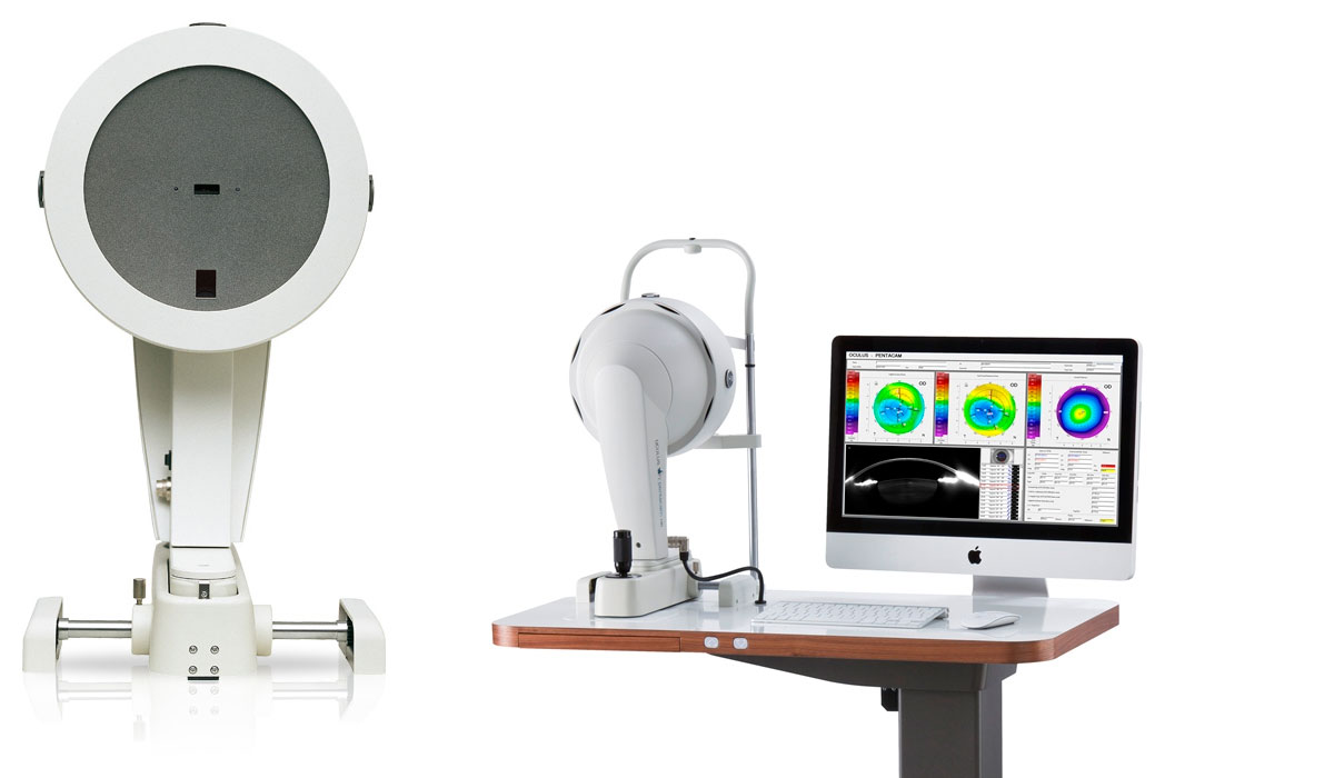
The Pentacam® is the only instrument on the market able to perform a precise and complete measurement and analysis of the centre of the cornea. It's a combined device consisting of a slit illumination system and a Scheimpflug camera which rotates around the eye.
The rotating measurement principle avoids measurement errors that would result from horizontal scanning. This contact-free, hygienic measurement process takes less than two seconds.
HOW DOES IT WORK?
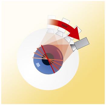
Pentacam works by illuminating a thin layer within the eye through its slit illumination system.
The cells in the eye are not entirely transparent and scatter in the slit light, in doing so they create a sectional image which is then photographed in side view by a camera.
The Scheimpflug camera captures an image of the illuminated plane from the anterior surface of the cornea right up to the posterior surface of the crystalline lens.
While swiveling around the eye, the slit-camera device generates a series of radially oriented images of the anterior eye chamber. In the subsequent analysis of the sectional images, tissue boundaries are detected and point clouds are assigned to the various tissue layers (anterior and posterior corneal surfaces, iris and crystalline lens).
The sectional images are saved, corrected in relation to a common reference point and then put together to create a three-dimensional model of the entire anterior eye chamber. This makes it possible to generate reproducible tomographic images of the anterior eye chamber in any desired plane.
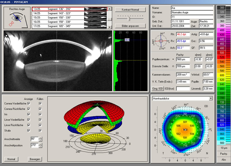
After correction for Scheimpflug distortion and light refraction, a tissue interfaces the exact location of image edge points in the eye by means of raytracing. Eye movements during image acquisition are captured by a second camera (pupil camera) and are also taken into account in the mathematical evaluation. This produces a set of three-dimensional measurement data which gives a precise geometric description of the anterior eye segment. This data in turn can be used to generate data on elevation, curvature, pachymetry, depth of the anterior eye chamber etc., in the well-known form of colour maps.
What is it for?
Within a mere second the Pentacam® supplies you with precise diagnostic data on the entire anterior eye segment. The degree of corneal or crystalline lens density is made visible by the light scattering properties of the respective media and is automatically quantified by the software. Measurement of the anterior and posterior corneal surfaces supplies the true total refractive power of the cornea. The data on the posterior surface support in Keratometry for toric IOL calculation and orientation as well as in the detection of early ectatic changes. The rotating scan supplies more data points in the center of the cornea. A supplementary pupil camera corrects for eye movements during the examination. Unlike conventional topography systems the Pentacam® measures true elevation data rather than just curvature values. Differences in brightness between individual layers provide an indication of any opacity, since transparent layers result in less slight scattering. This information can be used to assess cataracts.