CENTRAL SEROUS RETINOPATHY
Central serous retinopathy (CSR) is a recurring condition in which fluid accumulates in the fovea, the central region of the macula. It is an example of an
idiopathic disease which means that it has no known cause. However, it has been noted that CSR tends to be more common in male youths. In 95% of cases, the condition resolves, or heals, on its own with a full recovery of vision within 6-12 weeks. However, if recovery is prolonged, damage to the retina can occur which may result in mild to moderate vision loss. Laser treatments are sometimes used to speed up the natural healing process and minimize damage to the retina.
Even though the root cause of central serous retinopathy has yet to be identified, there are several risk factors associated with its development. The first is having an optic pit, a congenital “pit-like” formation on your optic nerve that may encourage the development of CSR. The second is progressive steroid use, which in some patients leads to the CSR, while another known trigger for the disease is an elevated stress level.
ASSOCIATION WITH OPTIC PIT
An optic pit is a defect of the optic nerve that can be associated with CSR, or blister formation in the central portion of the macula. In patients with CSR, it is important for an ophthalmologist to examine them for optic pit formations through careful diagnostic testing including Microperimetry, OCT scans, and Fluorescein Angiography.
IDIOPATHIC CENTRAL SEROUS RETINOPATHY
Idiopathic central serous retinopathy is used to refer to those patients who develop blistering conditions on the inside lining of the eye and for which the underlying cause is not known.
Central serous retinopathy is a condition in which blisters develop in the central foveal area of the macula. These blister formations are said to be recurrent and may occur bilaterally, meaning that both eyes are affected. When CSR develops, patients may notice substantial changes or distortion in their vision. Most commonly, patients will notice that objects in the eye that is affected are smaller than in the other eye.
In the early stages of this disease, there is no significant damage to the underlying retina if the CSR resolves itself quickly or if the patient is treated by an ophthalmologist. Patients with persistent central serous retinopathy are more prone to develop long-term, permanent retinal damage. Unfortunately, in patients with recurrent CSR, there may be progressive loss of sight each time there is new blistering in the retina. Accordingly, therapeutic interventions have been developed to reduce the duration of these blister formations in an attempt to maximize visual outcomes for these patients. Although idiopathic CSR is most commonly seen in young males, patients of all ages and gender have been diagnosed.
Older patients who develop idiopathic CSR may be at a greater risk of developing age related macular degeneration. Accordingly, patients who have CSR must be aware that further progressive damage may develop with time.
DETECTION & DIAGNOSIS
CSR is mainly a clinical diagnosis: that is, the doctor will make the diagnosis by listening to the medical symptoms described by the patient and examining the retina. However, additional diagnostic tests are usually performed to confirm the diagnosis and to determine what, if any, treatments may be necessary. The most important diagnostic test is Optical Coherence Tomography (OCT).
OCT testing will clearly show the fluid underneath the central retinal area and will allow your ophthalmologist to measure the amount of fluid present. This allows proper follow-up to determine how quickly the fluid is disappearing.
Another useful test diagnostic tool is the fluorescein angiography test, often used when there is some question about the exact cause of the fluid in the retina. In cases of CSR, the angiogram will often show a well-defined point of blood vessel leakage under the retina. The angiogram can also help in planning the optimal laser treatment for some cases of CSR.
Fundus Autofluorescence (FAF) testing is one of the latest diagnostic tests that has been very helpful in treating patients with CSR. It is particularly useful in showing evidence of previous episodes of CSR which may have caused mild damage to the retina.
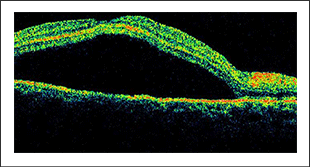
OCT image of retina affected by CSR.
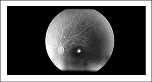
Fluorescein angiogram of CSR showing bright central spot of blood vessel leakage within the central retinal area.
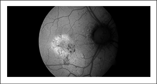
FAF image showing damage to cells within the retina from a previous episode of CSR.
PREVENTATION
VITAMINS
Vitamin supplementation has been proven effective in an age related eye diseases trial performed by the National Eye Institute in the United States for patients with age related macular degeneration (AMD).
Vitamin supplements reduce the rate of progression for specific subsets of these patients. Given that there are some similarities between patients with central serous retinopathy and those with age related macular degeneration, patients with central serous retinopathy may consider the use of vitamins supplementation to reduce the risk of disease progression.
STEROID EXPOSURE
There is a relationship between the use of steroids and the development of central serous retinopathy. While steroid usage is not found in all patients who develop CSR, it is recommended that patients who can avoid steroid use do so if they have had previous CSR. Steroids are found in nasal sprays, topical creams, and even some oral medications.
ANTIOXIDENTS
The use of antioxidant medication for patients who have macular degeneration has been well documented. Macular degeneration is a disease of the retinal pigment epithelium and is seen in a large number of the ageing population in North America. There is some relationship between disease mechanisms in patients who have central serous retinopathy and those who have age related macular degeneration. Accordingly, patients may consider the use of antioxidant medications such as vitamin supplementation to reduce the risk of progressive retinal damage. With that said, patients must understand that there is no proven evidence that this is helpful in patients with CSR.
STRESS REDUCTION
One of the common associations found in patients who have central serous retinopathy, apart from age and gender, is acute episodes of high stress. While many people undergo high episodes of stress, not all will develop central serous retinopathy. For those who develop central serous retinopathy, modifying social factors to reduce excessive stress is recommended, since this may reduce the development of recurrent CSR.
PROGNOSIS
Central Serous Retinopathy, or blister formations in the central macular area, tends to resolve spontaneously. However, the longer the duration of the blisters and the more recurrent they are, the more permanent visual loss will be.
Patients who develop CSR are at risk of developing age related macular degeneration as they age. Accordingly, patients who are found to have CSR, particularly in older age groups, may have further diagnostic interventions to ensure that there is no evidence of the wet, or advanced, form of age related macular degeneration. Clinical testing, including OCT, Fluorescein Angiography, and Microperimetry, can be essential in determining the risks of progression in these patients.
TREATMENT
SPONTANEOUS RESOLUTION
In approximately 95% of cases, CSR will resolve on its own without treatment and with full recovery of vision within 6-12 weeks from when the symptoms first developed. For this reason, in most cases of CSR, no treatment is required. For the first episode of CSR, observation is usually be recommended: the patient will be followed closely for a period of 1-3 months to ensure that the fluid underneath the retina is decreasing, indicating that it is likely to resolve completely within the usual 6-12 week period.
In certain instances, however, treatment may be recommended. The most common reason for recommending treatment is if the fluid takes longer than 12 weeks to dissipate, especially if vision is not improving. If fluid remains under the retina for an extended period of time, the risk of developing scar tissue under the retina greatly increases. It is this scar tissue that can cause permanent damage to vision. Another reason for recommending treatment is if a patient is experiencing recurrent episodes of CSR. Even when CSR resolves, up to 20% of patients will have a recurrence. Even though recurrences are often milder than the initial episode, when fluid accumulates repeatedly, the risk of scar tissue increases. An FAF diagnostic test is extremely helpful in showing evidence of previous episodes of CSR which may have caused scar tissue damage. Other reasons for recommending treatment is if there is an unusually large amount of fluid present, or for patients who require rapid improvement of vision, often for occupational reasons, and cannot wait 12 weeks for recovery.
Controlling the risk factors of CSR is also important. It may be recommended to stop the use of steroid nasal sprays or face creams, if possible. Stress management and blood pressure control may also be helpful. In very rare cases if an optic nerve pit is present, surgical options for correcting this may be
considered.
LASER TREATMENT
Treating central serous retinopathy involves monitoring its progress through diagnostic testing methods such as fluorescein angiography, OCT scans, and microperimetry. If a patient’s CSR does not heal fast enough, their ophthalmologist may use thermal or micropulse lasers to increase its natural rate of healing, thus minimizing the damage done to the central macular, or fovea.
Traditional management of CSR has been the use of high thermal energy lasers to treat focal points of leakage in the underlying layer of the retina known as the RPE. The goal of laser treatment is to reduce the amount of leakage by using laser photocoagulation to cauterize the leaking blood vessel. Although this is often successful, it also causes a scar in the retina that may cause vision problems.
More recently newer micropulse laser technology, which does not require clear definition of RPE focal leakage, has been used. This has the advantage of being able to stop the retinal leakage without causing any scarring to the retina and is the preferred method of treating CSR by the surgeons at OCC.
With the early use of laser treatments in patients with CSR, there can be earlier resolution of the central blister, reducing both the duration of the blister and the possibility of long-term visual damage. It is important to note that laser treatments do not reduce the risk of recurrence of the disease.
INTRAOCULAR INJECTION
Other treatments that have been used for CSR include intraocular injections of steroids or anti-VEGF drugs.
DEFINITIONS
FOVEA
The centra area of the retina onto which images observed by the eye are focused. Although it is less than a millimeter in diameter, it focuses all of the detailed visual input for the eye.
IDIOPATHIC
A disease or medical condition that has no known specific cause.
CSR
Central Serous Retinopathy
CONGENITAl
A medical condition that is present since birth, some of which are due to genetic abnormalities. Some of these conditions do not cause any significant medical problems, and are only found on a routine medical exam.
OPTICAL COHERENCE TOMOGRAPHY (OCT)
A diagnostic test that uses low intensity, invisible laser beams to measure the thickness of tissues within the retina. It produces a hi-definition, magnified image of the internal structures of the retina. The test is completely non-invasive and has no risk factors associated with it.
FUNDUS AUTOFLUORESCENCE (FAF)
A diagnostic test that uses a retina camera to illuminate the retina with a specific wavelength of light. This light causes certain molecules within the retina to glow, which is captured by the camera. The resulting image provides valuable information on the health status of the cells within the tissues of the retina. The test is completely non-invasive and risk-free.
LASER PHOTOCOAGULATION
A laser treatment in which the laser creates heat to cauterize abnormal blood vessels in the retina.
.png)
OCT image of the retina
.png)